
Figure 5.
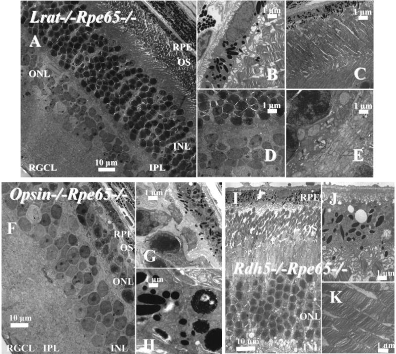
Montage of cross sections of the retinas of 2-month-old Lrat−/−Rpe65−/− mice analyzed by transmission electron microscopy. Panel A shows the cross section of the RPE and the photoreceptor cells. Panels B and C show higher magnification sections of the RPE and ROS (B, C) and the IPL (D, E). (F–H) Montage of cross sections of the retinas of 2-month-old opsin−/−Rpe65−/− mice analyzed by transmission electron microscopy. Panel F shows the cross section of the RPE and the photoreceptor cells. Panels G and H show higher magnification sections of the RPE and ROS, repectively. (I–K) Montage of cross sections of the retinas of 2-month-old Rdh5−/−Rpe65−/−mice analyzed by transmission electron microscopy. Panel I shows the cross section of the RPE and photoreceptor cells. Panels J and K show a higher magnification of the RPE and ROS. The sections were prepared as described in Experimental Procedures. The scale bar represents 1 or 10 μm as indicated.