
Figure 6.
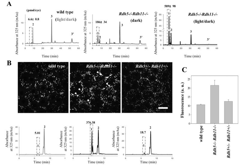
Light-dependent formation and storage of retinyl esters in eyes from Rdh5−/−Rdh11−/− mice. (A) Effect of dark or light rearing on the accumulation of 13-cis-retinyl esters in eyes from Rdh5−/−Rdh11−/− mice and wild-type controls. The dark-reared mice were exposed to no other light than dim red illumination. Peaks 1, 2, and 3/3′ represent cis-retinyl esters, all-trans-retinyl esters, and 11-cis-retinal oximes, respectively. The box represents cis-retinyl esters (>90% 13-cis-retinyl esters). A representative chromatogram is shown, and the average data from three mice are indicated with standard deviation above the ester peaks (mean ± SD). (B) Imaging of retinyl esters by two-photon microscopy (top row) and quantification of retinyl esters by HPLC (bottom row). Left column: Wild-type mice contained all-trans-retinyl esters in retinosomes (RESTs). Middle column: Rdh5−/−Rdh11−/− mice stored all-trans- and 13-cis-retinyl esters in retinosomes (white arrow). Retinosome fluorescence in Rdh5−/−Rdh11−/− mice is more intense than in wild-type mice (arrowheads). Right column: Distribution and quantity of all-trans-retinyl esters in eyes from Rdh5+/−Rdh+/− mice were similar to those of wild type. The box represents cis-retinyl esters (>90% 13-cis-retinyl esters). A representative chromatogram is shown, and the average data from three mice are indicated with standard deviation above the ester peaks (mean ± SD). (C) Quantification of fluorescence intensity measured by two-photon microscopy. Fluorescence intensity is higher in Rdh5−/−Rdh11−/− compared to wild-type and Rdh5+/−Rdh+/−mice (n = 3). The mean ± SD was indicated.