Figure 6.

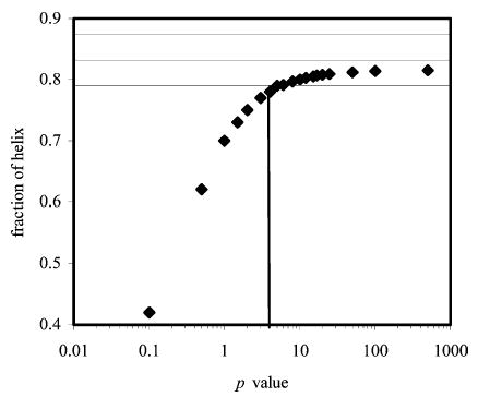
Plot showing the variation in the computed helix fraction of peptide KpS4 with the p value, as calculated using SCINT2 and doubly charged phosphoserine. Horizontal lines indicate the error range of the experimental values.

Plot showing the variation in the computed helix fraction of peptide KpS4 with the p value, as calculated using SCINT2 and doubly charged phosphoserine. Horizontal lines indicate the error range of the experimental values.