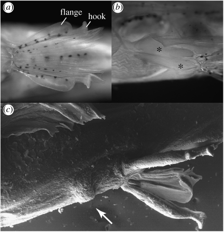
Figure 3.
(a) Paedocypris micromegethes, paratype male, ZRC 49869, 10.4 mm; pelvic fins, anteroventral view, showing hook and flange on anterior ray. (b) Paedocypris micromegethes, paratype, male, BMNH 2004.11.16.1-40, 10.9 mm, ventrolateral view on hypertrophied pelvic arrector and abductor muscles marked by asterisk symbols. (c) Paedocypris progenetica, paratype male, ZRC 43199, 8.5 mm, scanning electronic micrograph of pelvic region in ventrolateral view, arrow points to keratinized prepelvic knob.

