
Figure 1.
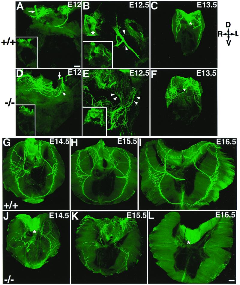
Aberrant innervation of the diaphragm muscle in mutant embryos from E12 to E16.5. All diaphragm images were shown as the top view. The orientation of the diaphragm is indicated in the upper right corner (D, dorsal; V, ventral; R, right; and L, left). In the controls (A), the intramuscular nerve trunk remained as a bundle, reached roughly at the middle of the dorsal diaphragm (arrow); the Inset is the low-power view. In the mutant, the phrenic nerves also reached the dorsal part of the diaphragm (D) but became aberrantly defasciculated (arrowhead). At E12.5, the phrenic nerve in the controls remained as a tight bundle with defined branches emanating perpendicularly from the main nerve trunk (arrows in B); the Inset is the low-power view. In the mutants, the phrenic nerve became more dramatically defasciculated (arrowheads in E) and distributed aberrantly across the entire dorsal surface of the diaphragm; the Inset is the low-power view. At E13.5, the phrenic nerves in the mutant (F) remained grossly defasciculated and aberrant, as compared with that in the control (C). At E14.5, the phrenic nerves in the controls (G) extended more toward dorsal and ventral portions of the diaphragm, innervating a ring of central band on the diaphragm, whereas the phrenic nerves in the mutants (J) were aberrantly distributed throughout the diaphragm and some nerves began to withdraw at this stage. By E15.5, only a small number of nerves remained in the diaphragm in the mutants (K). At E16.5, no phrenic nerve was observed in the entire diaphragm of the mutants [compare (I and L).] Note background staining associated with the esophagus (*) structure in some panels. [Bars = 100 μm (A, B, D, and E) and 200 μm (C, F, and G–L).]