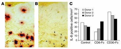
Figure 8. Increase in IL-8 expression in healthy skin mast cells after CD30-Fc stimulation of skin biopsies ex vivo.
Punch biopsies from 3 donors were subjected to submerged skin organ culture. Diluent control, 100 μg/ml CD6-Fc, or 100 μg/ml CD30-Fc was added to the culture. After 2 days in culture, skin specimens were processed for cryosections. (A) Sections were first enzyme-histochemically stained for tryptase. (B) After photography, sections were restained with a polyclonal antibody to IL-8. Arrows indicate cells that are positive for both tryptase and IL-8. (C) Number of IL-8 positive cells present after 2 days in culture was analyzed. Results are presented as number of IL-8 positive cells/mm2. Magnification, ×66.

