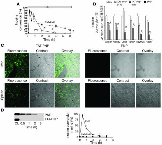
Figure 2. Transduction of TAT-PNP in PNP–/– mice.
TAT delivered active PNP into the tissues and prevented urinary excretion of the fused protein. PNP–/– mice (5–8 per group) were injected i.p. with 2.5 U/g body wt of TAT-PNP or PNP. Whole blood, tissue samples, and urine obtained from PNP–/– mice after injections were tested for the proteins’ presence and function after removal of membrane-bound proteins by washing with acidic buffer. PNP activity was determined by 14C-inosine conversion and is compared with PNP activity in samples obtained from normal control littermates (CL). (A) PNP activity in whole blood samples at the indicated time points after injections. (B) PNP activity in cell suspensions of various organs 4 and 8 hours after injections. (C) Detection of TAT-PNP intracellular transduction by confocal fluorescence microscopy in tissues 4 hours after injections. Sections were stained with rabbit anti-human PNP antibodies and secondary donkey Alexa 488–conjugated anti-rabbit antibody. Original magnification, ×200. (D) Urine samples were evaluated for the presence of recombinant proteins compared with 0.5 μg PNP. Western blot analysis with rabbit anti-PNP antibody and analysis of inosine conversion at the indicated time points after injections demonstrate that PNP, but not TAT-PNP, was excreted into the urine. Results (mean ± SD) are representative of 3 independent experiments. *P < 0.05.

