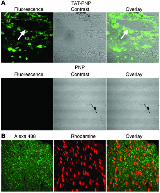
Figure 3. Presence of TAT-PNP in brain tissue from treated PNP–/– mice.
(A) Three-week-old PNP–/– mice were injected i.p. with 2.5 U/g body wt of TAT-PNP or nonfused PNP and sacrificed 4 hours later. Brain sections of mice were stained with rabbit anti-human PNP followed by Alexa 488–conjugated anti-rabbit antibodies and visualized by confocal microscopy. Arrow indicates blood vessel. Original magnification, ×200. (B) Brain sections obtained as described in A were also stained with mouse anti-neuronal nuclei antibodies. Rhodamine-conjugated anti-mouse was used as a secondary antibody. Original magnification, ×400. Images are representative of 3 independent experiments.

