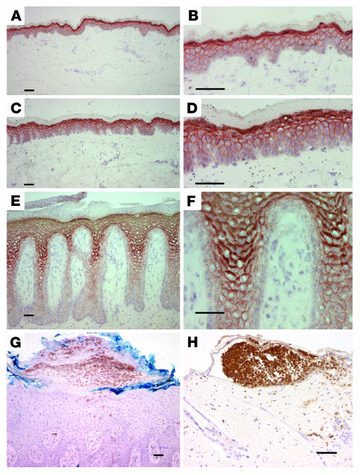
Figure 9. Prominent PKCα expression in the epidermis of psoriatic patients.
Frozen sections from normal (A and B), nonlesional (C and D), and psoriatic (E and F) epidermis were stained with monoclonal antibodies to PKCα and detected by immunoperoxidase. Results are typical of samples from 4 patients. Representative example of immunostaining for neutrophils in pustular psoriasis epidermis (G) and TPA-treated K5-PKCα mouse epidermis (H). PKCα staining in G (data not shown) was identical to E and F. Scale bars, 50 μm.

