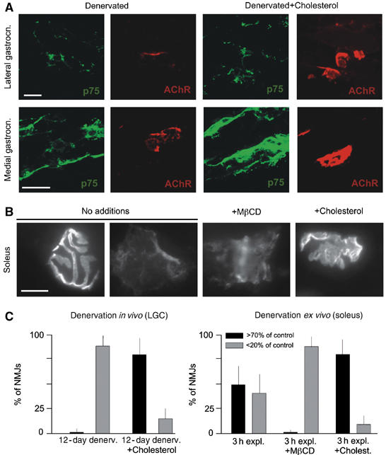
Figure 1.

Cholesterol stabilizes AChR clusters in denervated muscles. (A) Appearance of AChR clusters in two DeSyn muscles 12 days after denervation. Sciatic nerves were cut in 1 month mice; the absence of intact axons is confirmed by the expression of p75 in Schwann cells. Where indicated, cholesterol was applied daily, starting 5 days after denervation. The larger p75-positive area in the medial gastrocnemius panel with cholesterol is due to the particular plane of section, and does not reflect a systematic elevation of Schwann cell p75 immunofluorescence signals in denervated muscles treated with cholesterol. (B) Examples of AChR clusters (visualized by rhodamine-α-bungarotoxin; RITC-α-BT) in soleus nerve–muscle explants after 3 h in vitro. (C) Quantitative analysis of data as shown in (A) (left) and (B) (right). AChR labeling intensities (RITC-α-BT) were compared to controls; shown are fractions of NMJs with signal at least 70%, or less than 20% of control values. The 20 and 70% boundaries were selected to highlight the differences among the samples of these experiments. N=300 AChR clusters (from 3 mice each). Bars: 40 (A) and 20 μm (B).
