
Figure 6.
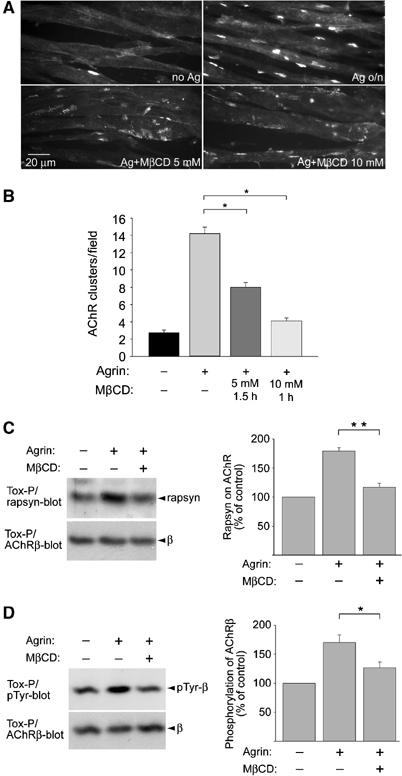
MβCD disrupts AChR clusters, AChR–rapsyn interaction and AChR β phosphorylation in C2C12 myotubes. (A) C2C12 myotubes where first treated overnight with agrin (Ag) to induce AChR clusters. MβCD was then added in the continued presence of agrin, causing AChR clusters to fragment and disappear, as revealed by rhodamine-α-BT staining. (B) Clusters of 5 μM minimal size were quantitated. (C, D) Cells were treated with agrin and MbCD (5 mM, 1.5 h) as in (A). AChRs were precipitated from cell lysates using biotin-α-BT (Tox-P). In immunoblots, AChR-associated rapsyn (C), phosphorylation of AChR β (D) and AChR β itself (C, D; whole protein-control) were detected and quantified by densitometric scanning (C, n=4; D, n=8). *P<0.05, **P<0.01, by two-tailed t-test.