
Figure 4.
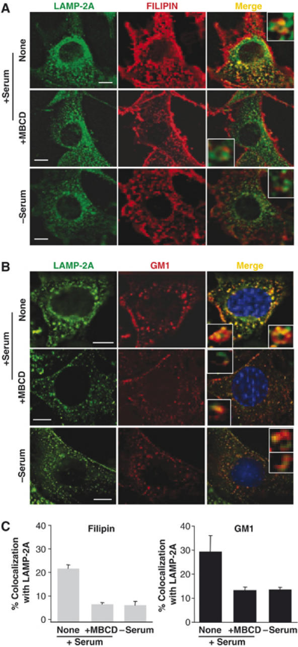
LAMP-2A localizes in cholesterol- and GM-1-enriched intracellular membrane domains. Mouse fibroblasts grown on coverslips in the presence (+) or absence (−) of serum were fixed and labeled with filipin (red) (A) or incubated with Texas-Red-cholera toxin B subunit to label GM-1-enriched membrane regions (red) and then fixed. (B) Both groups of cells were then subjected to indirect immunofluorescence for LAMP-2A (green). Where indicated, cells were treated with MBCD (25 mM) for 30 min before fixation. The merged images of both fluorophores are shown on the right. Insets show vesicles at higher magnification. Bar: 10 μm. The quantification of the colocalization of the two fluorophores is shown in (C). Values are expressed as the mean+s.e. of percentage of colocalization in 20–40 cells.