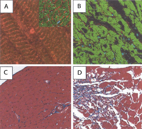
Figure 2.
ILK is required for cardiac muscle integrity. (A) Frozen section of heart from a wild-type FVB mouse that was incubated with an antibody specific for ILK, followed by a Cy3-conjugated secondary antibody. Note the expression of ILK (red) in the costa-meric regions of the murine cardiac myocytes. Inset shows the expression pattern in a cross-section of a wild-type heart, revealing ILK expression in the sarcolemma. Phalloidin stain appears green. (B) Frozen section of heart from an mckCRE ILKfl/fl mouse that was subjected to the same immunostaining protocol as in A. Note the absence of Cy3 signal in the heart tissue from this animal, indicating an absence of ILK expression. Cardiomyocytes from hearts of this genotype appear disaggregated following the loss of ILK. Hearts of this genetic combination were prepared from moribund animals exhibiting a dilated phenotype. (C,D) Hearts from control (C) and mckCRE ILKfl/fl (D) mice were sectioned and stained with trichrome stain. Note again the disaggregated tissue in the mckCRE ILKfl/fl genotype, in addition to large amounts of interstitial fibrosis (blue stain).

