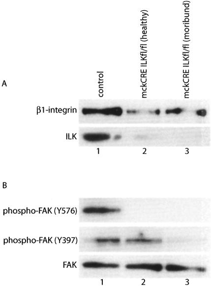
Figure 3.
Loss of ILK impacts β1-integrin/FAK signaling in mckCRE ILKfl/fl mice. (A) Pooled protein lysates from control (lane 1) and mckCRE ILKfl/fl (lanes 2,3) mice were subjected to immunoblot analysis, by probing with antibodies to β1-integrin (top panel) and ILK (bottom panel). Mice of the mckCRE ILKfl/fl genotype were selected at two time points: young, healthy animals not yet exhibiting evidence of cardiomyopathy (lane 2) and older mice showing evidence of morbidity due to cardiac failure (lane 3). Note that loss of ILK protein is accompanied by a corresponding reduction in β1-integrin protein levels. (B) Pooled lysates from A were subjected to immunoblot analysis for levels of FAK phospho-Tyr 576 (top panel) and phospho-Tyr 379 (bottom panel).

