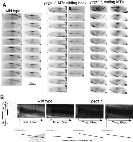
Figure 7.
Microtubule sliding did not account for the persistence of microtubules at cell tips. Regions of microtubules of the indicated GFP α-tubulin-expressing strains were marked by photobleaching the fluorescence in a microtubule bundle in a zone between the nucleus and the cell tip. (A) Images were taken in a single focal plane at 1-sec intervals. (B) Kymographs showing microtubules from the regions indicated in the cartoon on the left. The cartoon on the left of the kymographs indicates the region of the bleached micro-tubules used to generate the kymographic data. The cartoons below the kymographs summarize the rearward migration of the rear of the bleached zone and the growth of a microtubule through the bleached zone.

