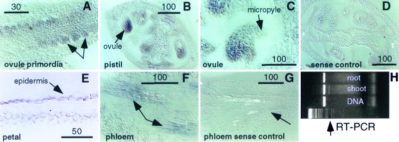
Figure 3.
Expression pattern of the FDH gene. (A–G) Light micrographs of wild-type floral tissues showing in situ RNA hybridization of the FDH antisense (A–C, E, and F) and sense (D and G) probes. Hybridization is detected in ovule primordia (arrows, A) as well as mature ovules (B and C). Petal epidermal cells (E) show a strong hybridization signal as does phloem tissue in vascular strands (arrows, F). Control sense strand hybridizations show no signal in the ovary (D) or vascular tissues (G). Reverse transcription–PCR confirms that FDH expression is limited to the shoot (H). Primers 3L and 2R (Materials and Methods) were used to specifically amplify a 256-bp genomic region that spans intron 2, which is 74 bp long. Maturation of the FDH mRNA results in the synthesis of a smaller 182-bp PCR product from the cDNA template (arrow). (Scale bars indicate magnification in μm.)

