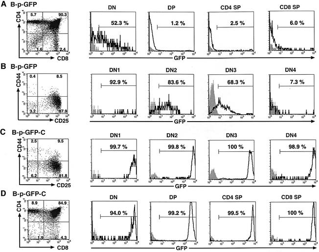
Fig. 5. B-p-GFP and B-p-GFP-C display distinct activities during T-cell differentiation. (A and D) The CD4/CD8 profiles in the adult thymus of B-p-GFP and B-p-GFP-C transgenic lines were revealed using antibodies to CD4 and CD8 conjugated to Cy-Chrome and R-PE, respectively. Each subpopulation was analyzed for GFP expression relative to its counterpart in a non-transgenic control. Histograms from control and transgenic populations are shown, and the percentage of the population falling within the positive gate is indicated. (B and C) Expression of B-p-GFP (B) and B-p-GFP-C (C) in the double-negative thymic precursors in the adult thymus. After gating out Lin+(TCRβ+/CD4+/CD8+/Mac1+/Gr1+/Ter119+/B220+/TCRγδ+/NK1.1+) cells, thymocytes were subdivided further into DN1–DN4 using antibodies to CD44 (APC) and CD25 (R-PE) antigens. The DN1–DN4 subpopulations were analyzed further for GFP expression in transgenic and non-transgenic controls. Histograms for GFP expression are shown, and the percentage of the population falling within the positive gate is indicated.

An official website of the United States government
Here's how you know
Official websites use .gov
A
.gov website belongs to an official
government organization in the United States.
Secure .gov websites use HTTPS
A lock (
) or https:// means you've safely
connected to the .gov website. Share sensitive
information only on official, secure websites.
