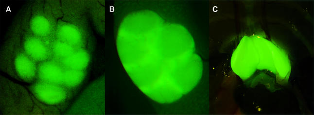
Fig. 8. Analysis of T and B cell-specific areas in B-p-GFP and B-p-GFP-C mice by fluorescence microscopy. (A) Peyer’s patch in the outer intestinal lining in the transgenic line B-p-GFP. Intense green staining marks B-cell follicles. (B) Peyer’s patch in the outer intestinal lining in the transgenic line B-p-GFP-C. The staining in the T-cell zone is more prominent than in the B-cell follicles. (C) Adult thymus of the transgenic line B-p-GFP-C.

An official website of the United States government
Here's how you know
Official websites use .gov
A
.gov website belongs to an official
government organization in the United States.
Secure .gov websites use HTTPS
A lock (
) or https:// means you've safely
connected to the .gov website. Share sensitive
information only on official, secure websites.
