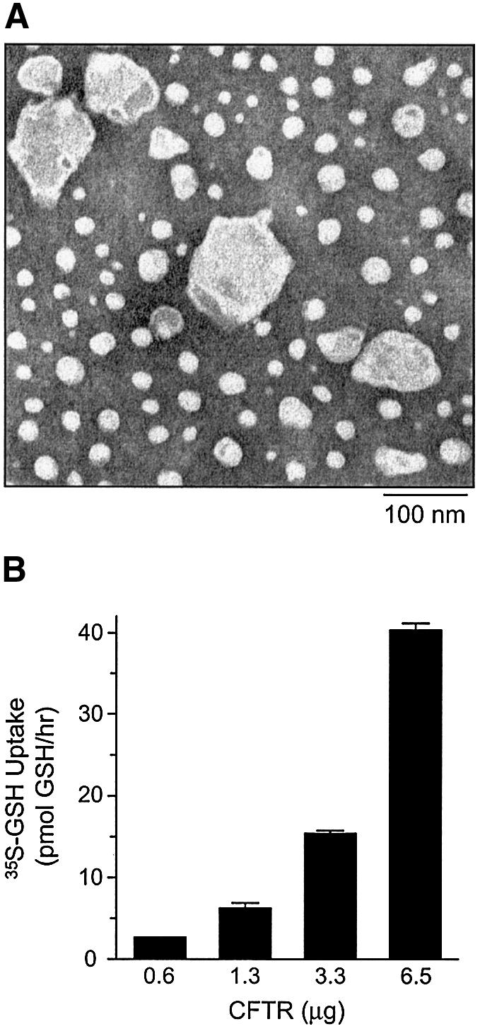
Fig. 5. GSH flux by purified and reconstituted CFTR protein. (A) Electron microscopy of purified and reconstituted wild-type CFTR. Proteoliposomes on carbon formvar-coated grids were stained with 2% uranyl acetate and visualized by negative staining. Magnification is ×150 000. Bar = 100 nm. (B) Increasing concentrations of reconstituted and phosphorylated wild-type CFTR protein (0.6–6.5 µg) were incubated with 20 nM [35S]GSH and 1 mM cold GSH in CFTR transport buffer, in the presence of MgAMP-PNP. Each value represents the mean of duplicate determinations.
