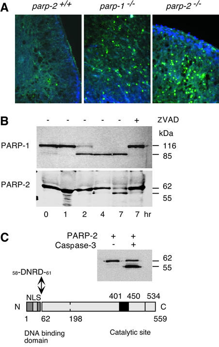
Fig. 4. (A) Fluorescent TUNEL stain of thymus 2 days after 6 Gy irradiation. Apoptotic cells are labeled in green, and cell nuclei are stained with DAPI. parp +/+ are sparsely stained whereas parp-1–/– and parp-2–/– show a strong green staining of apoptotic cells. (B) PARP-1 and PARP-2 cleavage kinetics after 100 µM VP16 treatment in HL-60 cells. Z-VAD-fmk inhibitor was used at 0.5 µg/µl and was added 1 h before VP16 treatment. For western blot, 105 cells were harvested at the indicated time, mixed with loading buffer and sonicated. Polyclonal antibodies anti-PARP-1 and anti-PARP-2 were used. (C) In vitro cleavage of purified mPARP-2 (2.5 µg) by active caspase-3 (8 U, Chemicon International) shows the same cleavage pattern in HL-60 cells after VP-16 treatment. The N-terminus of the 55 kDa band was micro sequenced and was found to be 58DNRD61, a consensus cleavage site for caspases 3/7.

An official website of the United States government
Here's how you know
Official websites use .gov
A
.gov website belongs to an official
government organization in the United States.
Secure .gov websites use HTTPS
A lock (
) or https:// means you've safely
connected to the .gov website. Share sensitive
information only on official, secure websites.
