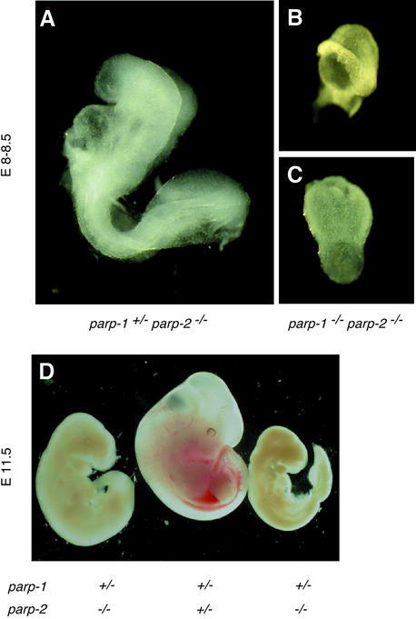
Fig. 5. External views of E8.0–8.5 (A–C) and E11.5 (D) embryos. (A) A normal parp-1+/–parp-2–/– embryo. (B and C) Retarded parp-1–/– parp-2–/– embryos. (D) Example of two parp-1+/–parp-2–/– female embryos that are phenotypically arrested at E9.5, and one normal parp-1+/–parp-2+/– female embryo at E11.5. The same magnifications were used in (A), (B) and (C).

An official website of the United States government
Here's how you know
Official websites use .gov
A
.gov website belongs to an official
government organization in the United States.
Secure .gov websites use HTTPS
A lock (
) or https:// means you've safely
connected to the .gov website. Share sensitive
information only on official, secure websites.
