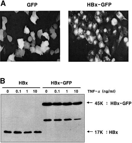
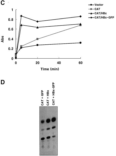
Fig. 1. Expression of HBx and HBx–GFP as functionally active forms. (A) Expression pattern of GFP and the HBx–GFP fusion protein. HepG2 cells were transfected with pEGFP and pEG-HBx-GFP plasmid. At 24 h post-transfection, cells were photographed by fluorescence microscopy (magnification ×200). (B) Expression of HBx and HBx–GFP analyzed by western blotting. HepG2 cells were transfected with pEG-HBx or pEG-HBx-GFP, and at 10 h post-transfection cells were treated with TNF-α for 20 h. Equal amounts of total cell lysates were analyzed by 12% SDS–PAGE. (C and D) Transcriptional transactivity of HBx and HBx–GFP in the CMV promoter-driven CAT reporter assay in HepG2 cells. At 24 h after co-transfection (0.5 µg each), whole-cell lysates were prepared and normalized for CAT assay with the calorimetric method (C) and TLC method (D).

An official website of the United States government
Here's how you know
Official websites use .gov
A
.gov website belongs to an official
government organization in the United States.
Secure .gov websites use HTTPS
A lock (
) or https:// means you've safely
connected to the .gov website. Share sensitive
information only on official, secure websites.

