Figure 2.
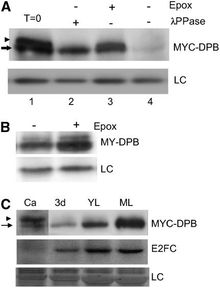
DPB Is Regulated by Phosphorylation and Proteolysis.
(A) Immunoblot analysis of MYC-DPBOE plant extracts treated with or without λ-phosphatase (λPPase). Where indicated, epoxomicin (Epox) was included in the reaction. The arrow indicates the MYC-DPB protein, and the arrowhead points to the phosphorylated MYC-DPB form. LC, loading control, corresponding to an unspecific cross-reacting protein; T=0, fresh extracts without any treatment.
(B) Immunoblot of protein extracts from MYC-DPBOE seedlings treated with (+) or without (−) the proteasome inhibitor epoxomicin. LC, loading control, corresponding to an unspecific cross-reacting protein.
(C) MYC-DPB and E2FC protein phosphorylation patterns at different stages of development. Ca, calli cells; 3d, 3-d-old seedlings; YL, young rosette leaves; ML, mature rosette leaves of 21-d-old plants. Top panel, anti-MYC; middle panel, anti-E2FC. Note that the Ca lane hybridized with anti-MYC was exposed five times longer than the others. LC, loading control, corresponding to the same gel stained with Coomassie blue. The arrow points to the MYC-DPB protein, and the arrowhead indicates phosphorylated MYC-DPB.

