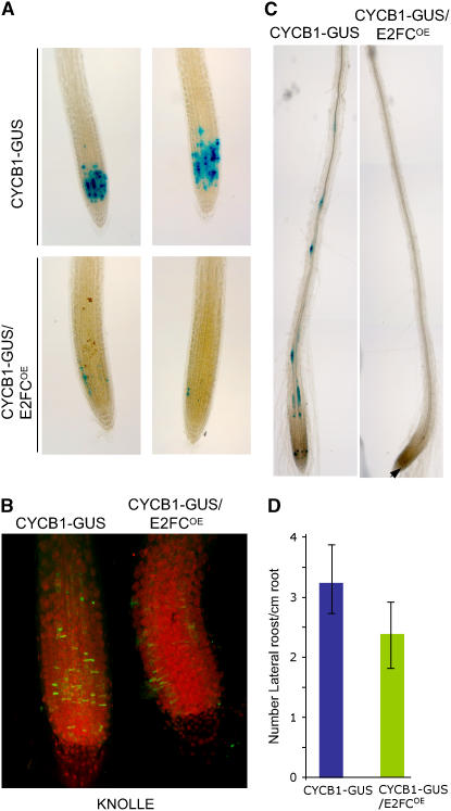
Figure 7.
Overexpression of E2FC Reduces the Level of CYCB1;1.
(A) GUS staining of CYCB1-GUS and CYCB1-GUS/E2FCOE roots of seedlings grown in MS medium for 7 d.
(B) KNOLLE protein immunolocalization (green) and propidium iodide staining (red) of CYCB1-GUS and CYCB1-GUS/E2FCOE roots. The image shows a stack of multiple confocal layers.
(C) GUS staining of roots of CYCB1-GUS and CYCB1-GUS/E2FCOE seedlings treated with auxin. The arrowhead indicates a GUS-stained cell in the meristem.
(D) Mean values of the numbers of LRP formed per centimeter of the main root in CYCB1-GUS and CYCB1-GUS/E2FCOE seedlings grown for 7 d in MS medium. Data are means of 40 seedlings in two independent experiments ± sd.

