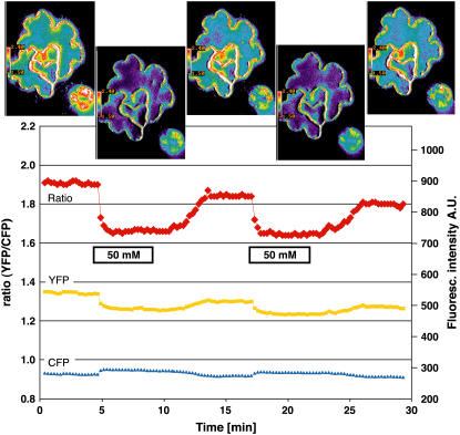
Figure 4.
Glucose-Induced FRET Changes in the Cytosol of Leaf Epidermal Cells.
The FRET sensor FLIPglu-600μΔ13 with an affinity of 600 μM for glucose in stably transformed rdr6-11 Arabidopsis plants responds in the epidermis to perfusion with 50 mM glucose. Quantitative data were derived by pixel-by-pixel integration of the ratiometric images. The scale at right gives fluorescence intensity in arbitrary units (A.U.) for the individual eCFP (480/30) and eYFP (535/40) emission channels; the scale at left gives the ratio of eYFP intensity divided by eCFP intensity. For each phase (plus/minus glucose), one ratiometric image with a pavement and a guard cell is shown.

