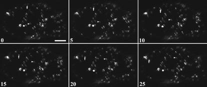
Figure 4.
Peroxisomal Motility in B. sinuspersici Chlorenchyma Cells Transiently Expressing GFP-MFP.
Time-lapse images of a GFP-MFP–expressing B. sinuspersici chlorenchyma cell. The movement of three peroxisomes was monitored for 25 s. One of these peroxisomes (stars) showed oscillatory movement over the entire series. Another peroxisome (arrowheads) remained fixed at a site for 5 s and then exhibited short-distance movement during the last 20 s. A third peroxisome (arrows) demonstrated continuous movement through the cytosol, covering a total distance of >40 μm. Numbers indicate elapsed time in seconds. Bar = 15 μm.

