Figure 5.
The Actin Cytoskeleton in Chlorenchyma Cells of B. sinuspersici and S. aralocaspica.
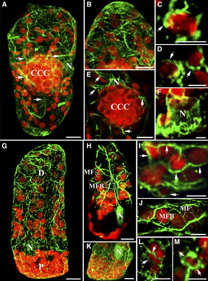
Immunofluorescence staining of actin demonstrates actin–chloroplast association in Bienertia and S. aralocaspica chlorenchyma cells. Actin filaments (green) were visualized with Oregon green–conjugated secondary antibody, and chloroplasts (red) were observed using their autofluorescence. Except for (A), (B), and (G), the images are single optical sections that demonstrate the direct interaction between actin filaments and chloroplasts. These images represent merged images of the dual channels that show their interaction. This is a representative result from at least five separate experiments with >50 cells observed. Arrows in (A) and (E) show the thick actin filament bundles connecting the CCC and the PCC. (G), (H), and (K) show that the nucleus (N) is also surrounded by actin filaments. Bars = 10 μm in (A), (B), (E), (G), (H), and (K) and 5 μm in (C), (D), (F), (I), (J), (L), and (M).
(A) and (G) Composite images (projections) of 30 optical 0.8-μm sections depicting the general actin filament patterns in Bienertia and S. aralocaspica chlorenchyma cells, respectively.
(B) Projection of a low-resolution image of the PCC showing the general distribution of the actin filaments.
(C) and (D) Single optical sections of high magnification of a region within the peripheral compartment demonstrating the close contact of actin filaments (arrows) with the chloroplasts.
(E) and (F) Single optical sections illustrating actin filaments surrounding and emanating from the nucleus (N).
(G) and (H) Projection (G) and single optical section (H) showing the two types of actin filaments: thick actin microfilament bundles (MFB) and thin actin microfilaments (MF) in S. aralocaspica.
(I) and (J) Single optical sections demonstrating the positioning of chloroplasts along the actin cables (arrows) by attaching to the thin actin filaments in the distal compartment.
(K) Optical section showing the actin filament pattern in the proximal compartment.
(L) and (M) Single optical sections of closeup images showing baskets of actin filaments (arrows) completely surrounding the chloroplasts.

