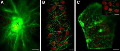
Figure 7.
Live Cell Localization of GFP Fusion Proteins.
B. sinuspersici chlorenchyma cells transiently expressing GFP chimeric proteins to actin binding protein (talin) or MAP4. Bars = 10 μm in (A) and (C) and 5 μm in (B) and inset in (C).
(A) Chlorenchyma cell transformed with GFP-talin showing thick actin filament bundles extending from the nucleus (N) and the CCC.
(B) Closeup image of a chlorenchyma cell transformed with GFP-talin showing actin filaments interacting with chloroplasts in the cortical region.
(C) Chlorenchyma cell transformed with GFP-MAP4 showing a dense network of microtubules in the cortical region. The inset shows an optical section through the cortical region of a GFP-MAP4–expressing cell showing both microtubules and chloroplasts.

