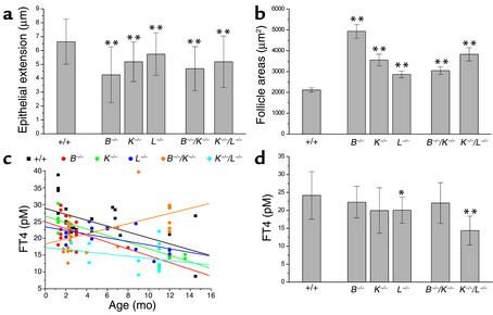
Figure 6.
Alterations in thyroid physiological parameters. (a and b) Morphometric analysis of epithelial extensions, i.e., cell heights (a), and follicle dimensions (b) of thyroids from WT and cathepsin-deficient mice. Extensions of thyroid epithelia were significantly reduced (a) whereas follicle areas were significantly increased (b) in mice of all cathepsin-deficient genotypes. (c and d) To determine whether flattening of epithelia reflects a reduction in the functional activities of thyroid glands, serum T4 levels were determined by a radiometric immunoassay. (c) The scatter graph indicates distribution of serum T4 levels plotted against mouse age. (d) Data from mice of all ages were analyzed. Serum T4 levels were significantly but slightly reduced in cathepsin L–/– mice (c and d, blue). A systemic defect in thyroid function was apparent from the very significantly reduced serum T4 levels in cathepsin K–/–/L–/– mice (c and d, cyan). Mean values ± SD are given in a and d, mean values ± SE in b. Lines represent linear regressions of the single data plotted against mouse age in c. *P < 0.05, **P < 0.01. In a and b, arbitrarily chosen follicles were analyzed; n = 162, 70, 71, 114, 150, and 58, respectively. In c and d, T4 levels of different animals were determined; n = 25, 12, 10, 14, 28, and 13, respectively.

