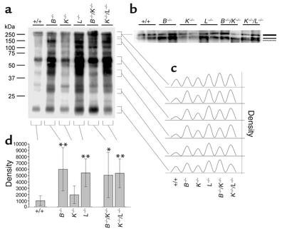
Figure 7.
Tg persistence in thyroids of mice with deficiencies in cathepsin B or L. Lysates of thyroids from mice of the indicated genotypes were normalized to equal amounts of protein, separated on 10% SDS gels, and transferred to nitrocellulose for subsequent incubation of the blots with antibodies against Tg. (a) A representative immunoblot. (b) The uppermost portion of another immunoblot demonstrates the expression of dimeric (thick line) and monomeric Tg (thin line) in WT and all cathepsin-deficient thyroids, as well as the appearance of a high–molecular weight Tg fragment (broken line), which was observed in the lysates of cathepsin K–/–/L–/– thyroids only. Molecular mass markers are indicated in the left margin. (c) Densitometric profiles of the indicated bands representing intact Tg and several of its degradation fragments. (d) Three different immunoblots with two lanes per genotype were evaluated densitometrically. Mean values ± SD of the densities of the lanes are given. Note that the amounts of immunolabeled Tg were upregulated about five- to sixfold in thyroids of cathepsin B– or L–deficient mice, whereas cathepsin K–/– thyroids contained only about twofold the amounts of Tg present in WTs (d).

