Figure 8.
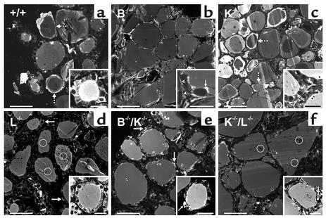
Multilayers of luminal Tg are lacking in thyroid follicles of cathepsin B–/–, L–/–, B–/–/K–/–, and K–/–/L–/– mice. Confocal fluorescence micrographs of cryosections of thyroid glands from WT mice (a) or cathepsin-deficient mice of the indicated genotypes (b–f) were immunolabeled with antibodies against Tg. As expected, Tg was detected within reticular structures and numerous intracellular vesicles (arrows), i.e., within compartments of the biosynthetic and of the endocytic route. Arrowheads indicate immunolabeled Tg in association with the ECM surrounding thyroid follicles. Open circles indicate non-immunolabeled inclusions of dead cells within the follicle lumina of cathepsin L–/– or K–/–/L–/– mice. In WT and cathepsin K–/– mice, immunolabeling revealed the characteristic multilayered appearance of Tg within the lumina of thyroid follicles (broken arrows), representing different states of Tg compaction. In contrast, Tg was homogeneously distributed within the follicle lumina of thyroids of cathepsin B–/–, L–/–, B–/–/K–/–, or K–/–/L–/– mice, indicating that deficiencies in either cathepsin B or cathepsin L resulted in the absence of differentially compacted luminal Tg. Bars: 50 μm; in insets: 20 μm.

