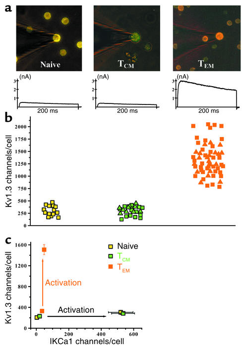
Figure 3.
Kv1.3highIKCa1low phenotype: an exclusive functional marker for activated effector memory T cells. (a) Fluorescent immunostained images of patch-clamped CD4 T cell subsets from control subjects showing naive CCR7+CD45RA+ (left), TCM CCR7+CD45RA–cells (middle), and TEM CCR7+CD45RA+cells (right, counterstained with antihuman CD4 mAb). Representative Kv1.3 currents from each cell type are shown below the respective images. (b) Kv1.3 channel number/cell in naive (yellow squares), TCM (MBP-stimulated, green squares; TT-stimulated, green triangles) and TEM (MBP-stimulated, orange squares; TT-stimulated, orange triangles) T cells. Activated naive CD4+ cells were identified in the peripheral blood of controls 48 hours following stimulation with anti-CD3 mAb. Activated TCM and TEM cells were identified in the same preparations of control TCLs (stimulated ten times with either MBP or TT) 48 hours after stimulation with the appropriate antigen. (c) Kv1.3 versus IKCa1 channel number per cell in the three populations before and after activation. Each data point is the mean ± SEM channel number in 20–50 cells.

