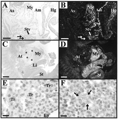
FIG. 1.
ΤβRIII mRNA expression within heart and liver during midgestation. Paired light- and dark-field photomicrographs of sections through mouse embryos, processed by in situ hybridization histochemistry by using an antisense cRNA probe for ΤβRIII. (A and B) A sagittal section through an E9.5 embryo shows ΤβRIII mRNA expression localized to the myocardium (My) of the developing ventricles and to the septum transversum (STv), amnion (Am), and hindgut (Hg). No expression is detected in the aortic sac (As) or presumptive hepatic endoderm (arrows). (C and D) A sagittal section through an E11.5 embryo shows high TβRIII mRNA expression in the myocardium of the ventricle (My) and atrium (At) but not in endocardial cushion tissue (*) of the heart. Strong expression is also now evident within the liver (Li) and stomach (St). (E) A light-field image of an E11.5 heart at high magnification shows silver grains localized over the myocytes within the trabeculae (Tr), with little expression within the myocytes immediately beneath the epicardium (Ep). (F) A similar image of E11.5 liver demonstrates strong TβRIII mRNA expression throughout the parenchyma. The arrows point to one of the islands of hepatocytes. Bars, 100 μm (A and B); 200 μm (C and D); 10 μm (E and F).

