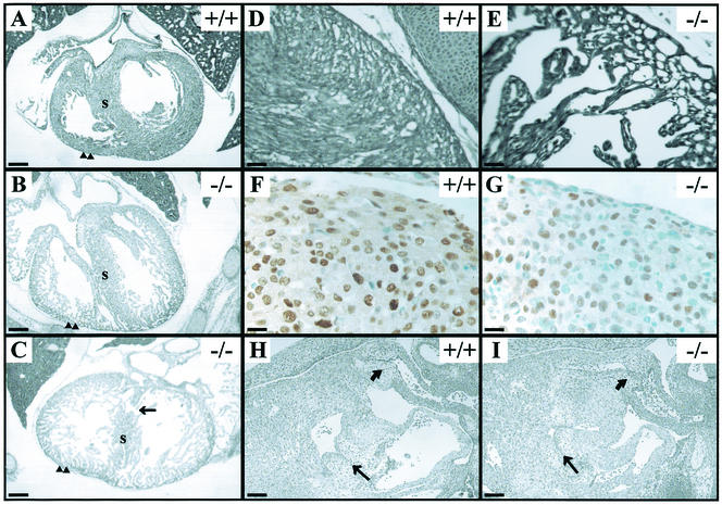
FIG. 5.
Heart defects in TβRIII−/− embryos. (A through C) Transverse sections through E16.5 hearts, near the level of the inlet valves, show thin, poorly compacted myocardial walls (arrowheads) and poorly formed septa in the TβRIII−/− hearts. (C) A severely affected heart, displaying a ventricular septal defect (arrow). (D and E) High-powered views of the compact zone in E18.5 hearts show an extreme example of compact zone thinness in the TβRIII−/− section. (F and G) PCNA immunostaining (brown) in E14.5 wild-type and knockout heart walls. Note the increase in the number of myocytes that do not express PCNA (blue cells) in TβRIII−/− heart. (H and I) Sagittal sections through E12.5 hearts show grossly normal atrioventricular (arrow) and outflow tract (curved arrow) endocardial cushion tissues in both wild-type and TβRIII−/− sections. Bars, 100 μm (A through C, H and I); 20 μm (D and E); 10 μm (F and G).

