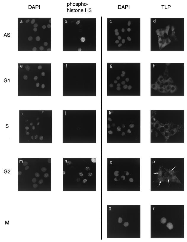
FIG. 3.
Cell cycle-specific nuclear translocation of cytoplasmic TLP. NIH 3T3 cells synchronistically staying at the G1/S boundary were released and cultured for 4, 8, and 12 h to prepare S-, G2-, and M-phase cell populations, respectively. For preparation of G1-phase cells, cells were synchronized at the early M phase and subsequently cultured for 4 h after release. AS, asynchronized cells. Cells were stained with DAPI, anti-phospho-histone H3, and anti-TLP antibody. Cell populations used for DAPI and anti-phospho-histone H3 staining were identical for the AS, G1, S, and G2 phases. Cells remaining in the M phase showed a homogeneous staining pattern with phospho-histone H3 (not shown). Among five cells remaining in the G2 phase, four nuclei (arrows) were stained with anti-TLP antibody (o and p). The microscopic images were taken at a magnification of ×640.

