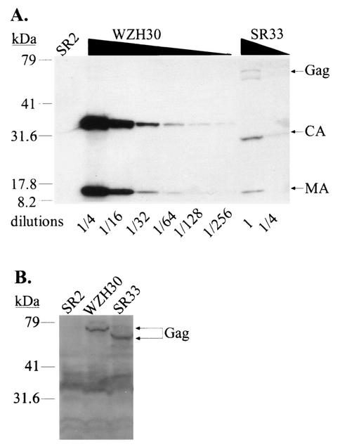
FIG. 3.
Characterization of pWZH30- and pSR33-transfected cells and virions derived from these cells. (A) Western analyses of cell-free virions. (B) Western analyses of cell lysates. Lanes labeled SR2 show results for supernatants or cell lysates from cells transfected with pSR2 and pSV-a-MLV-env; other lane labels specify supernatants or cell lysates from cells transfected with the indicated gag-pol expression construct plus pSR2 vector and pSV-a-MLV-env. Both anti-MLV CA and anti-MLV MA antibodies were used for the detection of virion lysates, whereas only anti-MLV CA antibody was used to detect cell lysates. The larger molecular-size bands (near the 79-kDa marker) detected in the SR33 virion lysates are probably caused by incomplete processing. A wide smear (between 30 to 40 kDa) present in the cell lysate analyses is presumably background created by nonspecific antibody binding. The migration of molecular size standards (in kilodaltons) and Gag, CA, and MA is shown.

