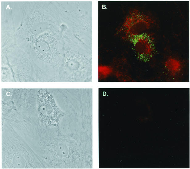
FIG. 3.
Colabeling of GFAP-positive cells with SIV p27. (A) Phase contrast of infected astrocytes plated on glass coverslips, infected with 50,000 cpm of SIV/17E-Br reverse transcriptase units, washed, and allowed to incubate for 1 week. (B) Immunofluorescence of astrocytes in A stained for viral p27 (fluorescein isothiocyanate; Dako) and GFAP (indocarbocyanine) (magnification, 400×). (C) Phase contrast of infected astrocytes (magnification, 400×). (D) Immunofluorescence of the astrocytes shown in C stained with isotype controls for both p27 and GFAP.

