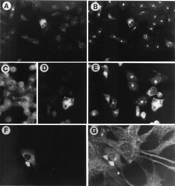
FIG. 3.
Pairs of micrographs of 143-tk− and Vero cells transfected with pwtgK-MTS and double stained with antibody to Myc (A, D, and F) and to giantin (B and E) or WGA (G). Anti-Myc antibody only stained the transfected cells, whereas the MAb to giantin and WGA stained all cells. In panels C to E, transfected cells were superinfected with R7032 24 h after transfection and fixed 10 h later. In panel C, cells were stained with anti-gD MAb to show that all cells in the culture were indeed infected. Panels A, D, and F show that anti-Myc staining was reticular and diffused to cytoplasm. This pattern overlaps with that of giantin in infected cells (E) but not in the uninfected cells (B and G). Panels A to E, 143-tk− cells; magnification, ×40. Panels F and G, Vero cells; magnification, ×63.

