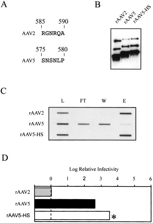
FIG. 7.
Modifying the heparin binding properties of AAV5. (A) Alignment of AAV2 amino acid residues 585 through 590 to residues predicted by amino acid alignment to be structurally equivalent in AAV5. (B) Western blot of iodixanol virus stocks. Equal volumes of virus were separated by SDS-10% polyacrylamide gel electrophoresis and analyzed by Western blotting using the B1 antibody. (C) Novel heparin binding properties of AAV5-HS. Heparin-agarose binding was performed as described in the legend for Fig. 2. See the legend for Fig. 2 for explanation of the abbreviations. (D) The log of the particle-to-infectivity ratio of the rAAV5 variants normalized to that of wild-type rAAV2, as described in the legend for Fig. 4.

