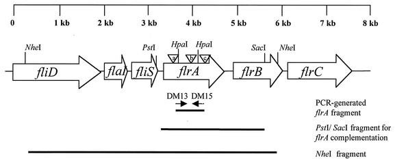
FIG. 1.
Schematic representation of the flrA locus in V. fischeri. Genes are designated by open boxes with arrows that indicate the direction of transcription. The internal fragment used to identify the flrA locus (using PCR primers DM13 and DM15), a PstI/SacI fragment inserted in pDM58 for flrA complementation, and the cloned NheI fragment are indicated by bold lines. Sequences upstream and downstream of the NheI sites were obtained from the V. fischeri genome sequencing project. The three triangles indicate the location of the TnflrA::Knr insertions (Table 1) in DM126 (a), DM127 (b), and DM128 (c). The sequence between the HpaI sites was removed to create the in-frame deletion strain DM159.

