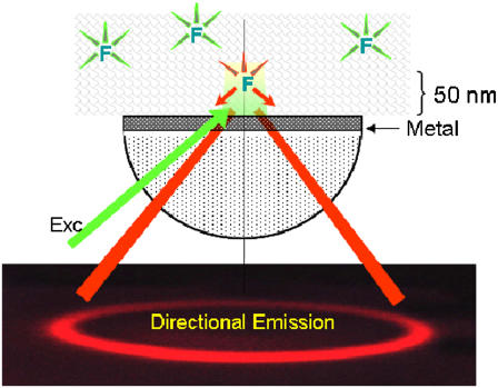
FIGURE 1.
Concept behind confocal SPCE microscope. The fluorophores are placed on a metal-coated coverslip and excited with green light at an SPR angle. The excitation energy couples to the surface plasmons and radiates to the glass prism (red) as a surface of a cone with half-angle equal to the SPCE angle. Metal can be a thin layer of Al (20 nm thick) or Ag or Au (50 nm thick). The picture of the directional emission is taken from a real experiment.

