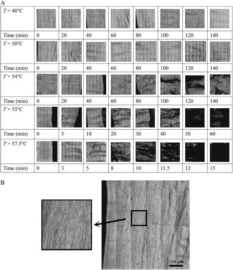
FIGURE 1.
(A) Second-harmonic generation images of rat tail tendon treated at different heating temperatures and times. Structural change at the fibril level of the rat tail tendon is visible in the SHG signal as the fibril is heated at or above 54°C. Very little structural change is observed when the heating temperature is below 54°C. The images are 660 μm × 660 μm in size, and each one is a montage of 36 individually scanned images. (B) A large image of the untreated 50°C specimen from A, with a magnified view showing the resolved tendon fibrils.

