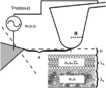
FIGURE 1.
Diagram of the tip-sample geometry used to generate a dielectrophoretic force on the sample; R is the radius of curvature (∼35 nm), d is the distance between the AFM tip and the counterelectrode, σ is conductivity, h is the height of the AFM tip over the membrane, C is the capacitance, ɛ is the permittivity, γ is the viscosity, and λ is the thickness of a given medium, with the subscripts m, c, and s referring to membrane, cytosol, and solution, respectively. The distance to the counterelectrode (25–100 μm) from the AFM tip is ∼25–100 μm.

