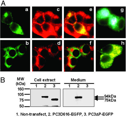
Figure 2.
The P domain is required for PC3 progression within the secretory pathway. (A) Analysis of subcellular localization by fluorescence microscopy. PC3D616–EGFP (a) and PC3ΔP–EGFP (b) expressed in βTC3 cells were observed as green. Endogenous insulin was identified by anti-insulin antibody (Cy3) (c and d) (red), and colocalization of EGFP-tagged proteins and endogenous insulin was observed as yellow in merged images (e and f). Golgi–enhanced cyan fluorescent protein and ER-enhanced yellow fluorescent protein were used as markers for Golgi (g) and ER (h), respectively. PC3D616–EGFP was predominantly localized in Golgi and secretory granules near the cell surface along with insulin, whereas PC3ΔP–EGFP was localized in the ER. (Magnification: ×800.) (B) Analysis of the expression and secretion of EGFP-tagged proteins by Western blotting using anti-GFP antibody. PC3D616–EGFP was detected as a 94-kDa band in both cell extract and medium. PC3ΔP–EGFP was detected in cell extract as a 75-kDa band, but was not detected in the medium.

