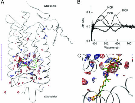
Fig. 1.
Conversion from RHO to LUMI in a 3D crystal. (A) Difference electron densities calculated from x-ray diffraction data at 160 K. Three purple double arrows indicate the slab sections shown in Fig. 3 A–C. (B) Spectral changes observed upon conversion from RHO to BATHO (100 K) and to LUMI (130 K, 140 K) in a 3D crystal. These spectra were obtained after subtraction of the ground-state spectrum. (C) Expanded view of the difference electron densities around the retinal. In A and C, positive and negative electron densities are shown in blue and red, respectively, on the ground-state α-carbon polypeptide chain of RHO. The maps are contoured to 3.5σ level for one of the two molecules in the crystallographic asymmetric unit. The chromophores (11-cis-retinal + Lys-296) of RHO and LUMI are in green and orange, respectively. Figs. 1 and 3 were prepared with SPDBV (15).

