Fig. 5.
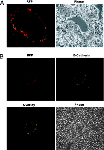
MET in lung metastasis. (A) Fluorescence and phase-contrast images of a lung section from animals harboring AT3-RIIIcI2 tumors (×200 magnification). Fluorescent cells are situated around a blood vessel (confirmed by hematoxylin/eosin staining; data not shown). (B) Example of a section from lungs of an animal bearing an AT3-RIIIcI2 tumor shows that the great majority of red fluorescent metastatic cells also stained positive for E-cadherin. Images were acquired at ×200 magnification.

