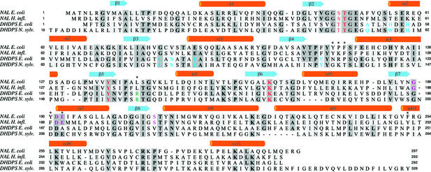
Figure 2.
Sequence alignment of NAL from E. coli, NAL from H. influenzae, DHDPS from E. coli, and DHDPS from N. sylvestris. Numbering for the latter is as in ref. 15. Secondary structural elements as found in NAL from E. coli are shown above the alignment. Residues identical in at least three of the four sequences are highlighted in dark gray, and similar residues are highlighted in light gray. Color coding: blue, residues forming l-lysine binding pocket in DHDPS (14, 15); red, residues involved in binding pyruvate or aldol condensation/cleavage reaction; magenta, residues specifically interacting with the aldol acceptor moiety of Neu5Ac (9, 37); green, residue binding the carboxylate group of l-ASA (14). Residues in NAL E. coli that were targeted for mutagenesis are marked with an asterisk. The alignment was done by using the program CLUSTALW (38) followed by manual editing.

