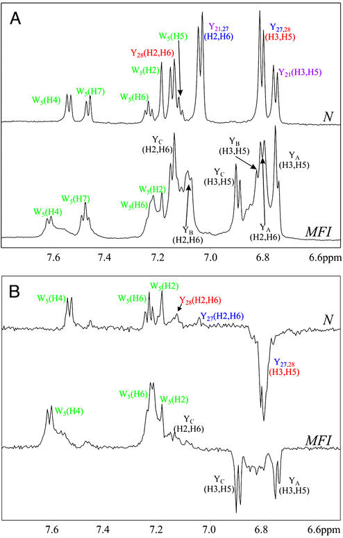
Figure 2.
1H NMR (600 MHz) (A) and photo-CIDNP (B) spectra of the purified native state (N) and of the MFI (0.3 mM protein/0.2 mM flavin mononucleotide, pH* 7, in 2H2O at 25°C). NMR and photo-CIDNP spectra were recorded with 512 and 16 scans, respectively. The assignments of the N-state tyrosine resonances are taken from Lu et al. (8). The tyrosine resonances of the MFI species, labeled YA, YB, and YC, have not been assigned to specific residues. Attempts to measure the spectra of the fully reduced protein at pH 7 were thwarted by the rapid formation of disulfide species (Fig. 1A). (pH* is the measured pH of a 2H2O solution, uncorrected for the deuterium isotope effect.)

