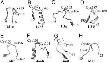
Figure 4.
Schematic view of the vicinal disulfide bonds, which have been structurally characterized. (A) 1dlo: J-Atracotoxin (H. versuta), C13-C14 (26); (B) 1eh5: palmitoyl protein thioesterase I (B. taurus), C45-C46 (32); (C) 1flg: ethanol dehydrogenase (P. aeruginosa), C105-C106 (30); (D) 1i9b acetylcholine-binding protein (L. stagnalis), C187-C188 (31); (E) 1obr: carboxypeptidase T (T. vulgaris), C155-C156 (34); (F) 4aah: methanol dehydrogenase (M. extorquens), C103-C104 (28); (G) 1m4f: hepcidin-25 (H. sapiens), C13-C14 (33); (H) Computer model of MFI, C17-C18 (see text).

