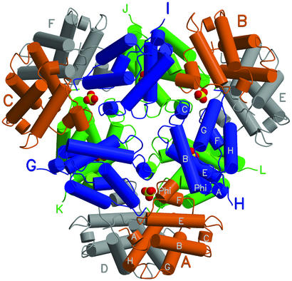
Figure 1.
A view of the assembled trHbO dodecamer down the local 3-fold axis direction. α-Helices are represented by cylinders (white labels in two subunits); heme groups have been omitted for clarity. The upper triangle six subunits are A, B, C, G, H, and I (orange and blue, large color-coded labels). The lower triangular assembly (green and gray subunits) is labeled with smaller lettering. Six sulfate anions are shown as yellow/red space filling models (partly hidden). The dodecamer's local 2-fold axes stretch from the center to the periphery of the assembly, through the six sulfate anions shown. All figures were drawn with MOLSCRIPT and RASTER3D (36, 37).

